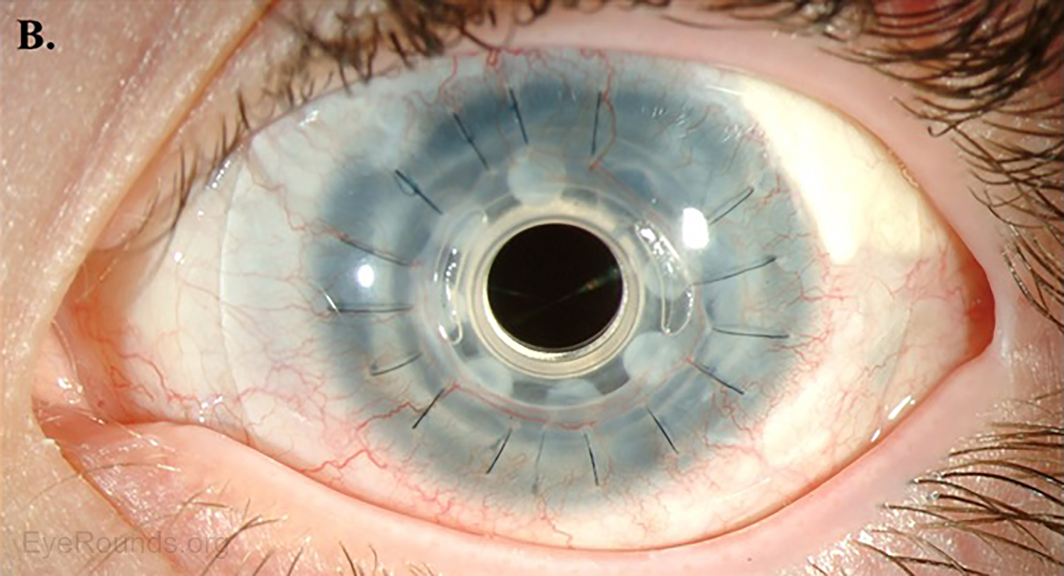
Slit Lamp Examination Of Cornea - Representative Slit Lamp Biomicroscopic Photograph Of Cornea In Cases 1 Representative Slit Lamp Biomicroscopic Photograph Of Cornea In Cases 1 And 2 Case 1 A

Last update images today Slit Lamp Examination Of Cornea

Slit Lamp Examination Ento Key   C06f239
Slit Lamp Examination Ento Key C06f239
Cornea And Anterior Eye Assessment With Slit Lamp Biomicroscopy   2 Figure1 1
Cornea And Anterior Eye Assessment With Slit Lamp Biomicroscopy 2 Figure1 1
A Slit Lamp Biomicroscopy Imaging Of The Cornea Of This Patient   A Slit Lamp Biomicroscopy Imaging Of The Cornea Of This Patient Showing An Irregular
A Slit Lamp Biomicroscopy Imaging Of The Cornea Of This Patient A Slit Lamp Biomicroscopy Imaging Of The Cornea Of This Patient Showing An Irregular
Slit Lamp Exam Corneal Abrasion   5081d90d2e947
Slit Lamp Exam Corneal Abrasion 5081d90d2e947
Figure Slit Lamp Image Of Cornea Iris And Lens StatPearls NCBI   Cornea
Figure Slit Lamp Image Of Cornea Iris And Lens StatPearls NCBI Cornea
Slit Lamp Exam Corneal Abrasion   Sddefault
Slit Lamp Exam Corneal Abrasion Sddefault
Slit Lamp Examination Of The Patient A Slit Lamp Examination   Slit Lamp Examination Of The Patient A Slit Lamp Examination Revealed Oval Shaped
Slit Lamp Examination Of The Patient A Slit Lamp Examination Slit Lamp Examination Of The Patient A Slit Lamp Examination Revealed Oval Shaped
Corneal Imaging An Introduction   Fig13b LRG
Corneal Imaging An Introduction Fig13b LRG
Slit Lamp Examination Performance At 10 Days After Surgery Moderate   Slit Lamp Examination Performance At 10 Days After Surgery Moderate Peripheral Corneal
Slit Lamp Examination Performance At 10 Days After Surgery Moderate Slit Lamp Examination Performance At 10 Days After Surgery Moderate Peripheral Corneal
Representative Slit Lamp Biomicroscopic Photograph Of Cornea In Cases 1   Representative Slit Lamp Biomicroscopic Photograph Of Cornea In Cases 1 And 2 Case 1 A
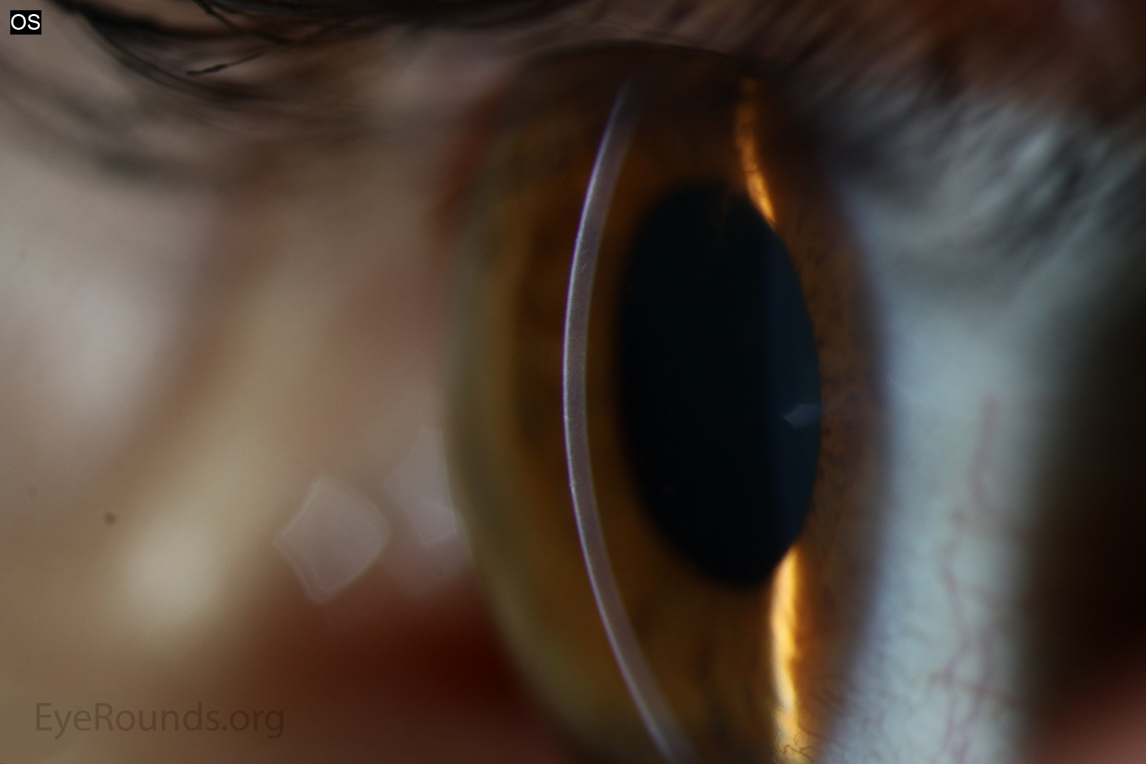
Representative Slit Lamp Biomicroscopic Photograph Of Cornea In Cases 1 Representative Slit Lamp Biomicroscopic Photograph Of Cornea In Cases 1 And 2 Case 1 A
Slit Lamp Exam Corneal Abrasion   Big 547454f18b185
Slit Lamp Exam Corneal Abrasion Big 547454f18b185
A To C A Corneal Slit Lamp Photograph Of Patient 1 A 30 Year Old   A To C A Corneal Slit Lamp Photograph Of Patient 1 A 30 Year Old Female With Corneal.ppm
A To C A Corneal Slit Lamp Photograph Of Patient 1 A 30 Year Old A To C A Corneal Slit Lamp Photograph Of Patient 1 A 30 Year Old Female With Corneal.ppm
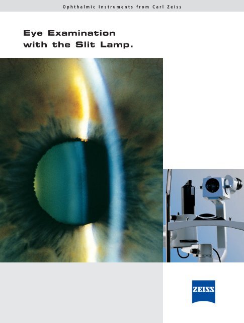
Eye Examination With The Slit Lamp Carl Zeiss Inc   Eye Examination With The Slit Lamp Carl Zeiss Inc
Eye Examination With The Slit Lamp Carl Zeiss Inc Eye Examination With The Slit Lamp Carl Zeiss Inc
Slit Lamp Examination Ento Key   C06f237
Slit Lamp Examination Ento Key C06f237
A Slit Lamp Photograph Of The Cornea 1 Minute After The Applanation   A Slit Lamp Photograph Of The Cornea 1 Minute After The Applanation With The Biopore
A Slit Lamp Photograph Of The Cornea 1 Minute After The Applanation A Slit Lamp Photograph Of The Cornea 1 Minute After The Applanation With The Biopore
Slit Lamp Photograph Zoomed In On The Cross Section Of The Cornea And   Slit Lamp Photograph Zoomed In On The Cross Section Of The Cornea And The Retained
Slit Lamp Photograph Zoomed In On The Cross Section Of The Cornea And Slit Lamp Photograph Zoomed In On The Cross Section Of The Cornea And The Retained
Slit Lamp Exam Corneal Abrasion   Initial Slit Lamp Image Showing A Fluorescein Stained Deep Paracentral Corneal Ulcer With
Slit Lamp Exam Corneal Abrasion Initial Slit Lamp Image Showing A Fluorescein Stained Deep Paracentral Corneal Ulcer With
Slit Lamp To Diagnose The Eyes And Cornea Slit Lamp For   Slit Lamp To Diagnose Eyes Cornea Slit Lamp Ophthalmologist Examination Eye Cornea Patient 279929900
Slit Lamp To Diagnose The Eyes And Cornea Slit Lamp For Slit Lamp To Diagnose Eyes Cornea Slit Lamp Ophthalmologist Examination Eye Cornea Patient 279929900
Slit Lamp Photograph Of The Cornea Before And After Corneal   Slit Lamp Photograph Of The Cornea Before And After Corneal Neurotization At Sequential
Slit Lamp Photograph Of The Cornea Before And After Corneal Slit Lamp Photograph Of The Cornea Before And After Corneal Neurotization At Sequential
A Clinical Slit Lamp Photograph Of The Affected Cornea At 6 Week Post   A Clinical Slit Lamp Photograph Of The Affected Cornea At 6 Week Post Initial
A Clinical Slit Lamp Photograph Of The Affected Cornea At 6 Week Post A Clinical Slit Lamp Photograph Of The Affected Cornea At 6 Week Post Initial
Slit Lamp Examination Photos On The Twenty Fifth Day After The Second   Slit Lamp Examination Photos On The Twenty Fifth Day After The Second Dose Of COVID 19
Slit Lamp Examination Photos On The Twenty Fifth Day After The Second Slit Lamp Examination Photos On The Twenty Fifth Day After The Second Dose Of COVID 19
A Slit Lamp Photograph Of The Cornea 1 Minute After The Applanation   A Slit Lamp Photograph Of The Cornea 1 Minute After The Applanation With The Biopore
A Slit Lamp Photograph Of The Cornea 1 Minute After The Applanation A Slit Lamp Photograph Of The Cornea 1 Minute After The Applanation With The Biopore
Ophthalmology Dx Blurred Sight For Sore Eye Ophthalmology Advisor   0621 Slitlamp 1 860x582
Ophthalmology Dx Blurred Sight For Sore Eye Ophthalmology Advisor 0621 Slitlamp 1 860x582
A Slit Lamp Photograph Of The Cornea 1 Minute After The Applanation   A Slit Lamp Photograph Of The Cornea 1 Minute After The Applanation With The Biopore
A Slit Lamp Photograph Of The Cornea 1 Minute After The Applanation A Slit Lamp Photograph Of The Cornea 1 Minute After The Applanation With The Biopore
Indian Journal Of Ophthalmology   SocialThumb.02223307 201866020 00005.T1 5
Indian Journal Of Ophthalmology SocialThumb.02223307 201866020 00005.T1 5
Slit Lamp Biomicroscopy Images Of The Corneas Of All 10 Patients At 4   Slit Lamp Biomicroscopy Images Of The Corneas Of All 10 Patients At 4 Years After Q640
Slit Lamp Biomicroscopy Images Of The Corneas Of All 10 Patients At 4 Slit Lamp Biomicroscopy Images Of The Corneas Of All 10 Patients At 4 Years After Q640
Slit Lamp Biomicroscopy Of Corneas At 3 Months After Hyperopic SMILE   Slit Lamp Biomicroscopy Of Corneas At 3 Months After Hyperopic SMILE Th400 D A Q640
Slit Lamp Biomicroscopy Of Corneas At 3 Months After Hyperopic SMILE Slit Lamp Biomicroscopy Of Corneas At 3 Months After Hyperopic SMILE Th400 D A Q640
Slit Lamp Photograph Of The Left Eye Showing Clear Cornea With A   Slit Lamp Photograph Of The Left Eye Showing Clear Cornea With A Superotemporal Aurolab
Slit Lamp Photograph Of The Left Eye Showing Clear Cornea With A Slit Lamp Photograph Of The Left Eye Showing Clear Cornea With A Superotemporal Aurolab
Lecture Using The Slit Lamp Microscope To Visualize The Ocular   Maxresdefault
Lecture Using The Slit Lamp Microscope To Visualize The Ocular Maxresdefault
A Case 2 At Initial Examination A Slit Lamp Photograph Showed A   A Case 1 At Initial Examination A Slit Lamp Photograph Showed A Peripheral Corneal Q640
A Case 2 At Initial Examination A Slit Lamp Photograph Showed A A Case 1 At Initial Examination A Slit Lamp Photograph Showed A Peripheral Corneal Q640
Slit Lamp Photography Of Normal Cornea A And Subepithelial   Slit Lamp Photography Of Normal Cornea A And Subepithelial Infiltrates In Case 1 B
Slit Lamp Photography Of Normal Cornea A And Subepithelial Slit Lamp Photography Of Normal Cornea A And Subepithelial Infiltrates In Case 1 B
Atlas Entry Normal Cornea   Normal Cornea LRG
Atlas Entry Normal Cornea Normal Cornea LRG
Understanding The Slit Lamp   Understanding The Slit Lamp 33 728
Understanding The Slit Lamp Understanding The Slit Lamp 33 728

https i2 wp com entokey com wp content uploads 2022 11 c06f239 jpg - Slit Lamp Examination Ento Key C06f239 https www researchgate net publication 321791604 figure fig1 AS 599537878048768 1519952377254 Initial slit lamp image showing a fluorescein stained deep paracentral corneal ulcer with png - Slit Lamp Exam Corneal Abrasion Initial Slit Lamp Image Showing A Fluorescein Stained Deep Paracentral Corneal Ulcer With

https www researchgate net profile Marco Lombardo 6 publication 306128774 figure fig2 AS 1086750420152335 1636112899873 A Slit lamp photograph of the cornea 1 minute after the applanation with the Biopore jpg - A Slit Lamp Photograph Of The Cornea 1 Minute After The Applanation A Slit Lamp Photograph Of The Cornea 1 Minute After The Applanation With The Biopore https media us amboss com media views 5081d90d2e947 jpg - Slit Lamp Exam Corneal Abrasion 5081d90d2e947 https www ncbi nlm nih gov books NBK539690 bin Cornea jpg - Figure Slit Lamp Image Of Cornea Iris And Lens StatPearls NCBI Cornea

https www researchgate net publication 260090503 figure fig1 AS 601778735292416 1520486639174 A Case 1 at initial examination a slit lamp photograph showed a peripheral corneal Q640 jpg - A Case 2 At Initial Examination A Slit Lamp Photograph Showed A A Case 1 At Initial Examination A Slit Lamp Photograph Showed A Peripheral Corneal Q640 https www researchgate net publication 361483442 figure fig1 AS 11431281094731778 1667549259642 Slit lamp examination photos on the twenty fifth day after the second dose of COVID 19 png - Slit Lamp Examination Photos On The Twenty Fifth Day After The Second Slit Lamp Examination Photos On The Twenty Fifth Day After The Second Dose Of COVID 19

https www ophthalmologyadvisor com wp content uploads sites 24 2021 05 0621 slitlamp 1 860x582 jpeg - slit edema corneal ophthalmology dx blurred sight sore Ophthalmology Dx Blurred Sight For Sore Eye Ophthalmology Advisor 0621 Slitlamp 1 860x582

https www researchgate net profile May Griffith publication 259490107 figure fig1 AS 267421160308786 1440769581581 Slit lamp biomicroscopy images of the corneas of all 10 patients at 4 years after Q640 jpg - Slit Lamp Biomicroscopy Images Of The Corneas Of All 10 Patients At 4 Slit Lamp Biomicroscopy Images Of The Corneas Of All 10 Patients At 4 Years After Q640 https www researchgate net publication 352722428 figure fig1 AS 1038409455906817 1624587515791 Slit lamp photograph of the left eye showing clear cornea with a superotemporal Aurolab png - Slit Lamp Photograph Of The Left Eye Showing Clear Cornea With A Slit Lamp Photograph Of The Left Eye Showing Clear Cornea With A Superotemporal Aurolab

https www researchgate net publication 363169055 figure fig3 AS 11431281082593742 1662051674209 Slit lamp photograph zoomed in on the cross section of the cornea and the retained jpg - Slit Lamp Photograph Zoomed In On The Cross Section Of The Cornea And Slit Lamp Photograph Zoomed In On The Cross Section Of The Cornea And The Retained https image slidesharecdn com understandingtheslitlamp 120722183047 phpapp02 95 understanding the slit lamp 33 728 jpg - slit cornea Understanding The Slit Lamp Understanding The Slit Lamp 33 728

https www researchgate net publication 324224732 figure fig1 AS 612091920654336 1522945494049 Slit lamp biomicroscopy of corneas at 3 months after hyperopic SMILE th400 D A Q640 jpg - Slit Lamp Biomicroscopy Of Corneas At 3 Months After Hyperopic SMILE Slit Lamp Biomicroscopy Of Corneas At 3 Months After Hyperopic SMILE Th400 D A Q640 https www researchgate net profile Marco Lombardo 6 publication 306128774 figure fig6 AS 1086750424338432 1636112900033 A Slit lamp photograph of the cornea 1 minute after the applanation with the Biopore jpg - A Slit Lamp Photograph Of The Cornea 1 Minute After The Applanation A Slit Lamp Photograph Of The Cornea 1 Minute After The Applanation With The Biopore https www ncbi nlm nih gov books NBK539690 bin Cornea jpg - Figure Slit Lamp Image Of Cornea Iris And Lens StatPearls NCBI Cornea

https i ytimg com vi 1E sEhy9tBo maxresdefault jpg - Lecture Using The Slit Lamp Microscope To Visualize The Ocular Maxresdefault https www researchgate net publication 361880985 figure fig1 AS 1180697335218176 1658511589092 Representative slit lamp biomicroscopic photograph of cornea in cases 1 and 2 Case 1 a png - Representative Slit Lamp Biomicroscopic Photograph Of Cornea In Cases 1 Representative Slit Lamp Biomicroscopic Photograph Of Cornea In Cases 1 And 2 Case 1 A

https www researchgate net profile Trevor Sherwin publication 6372419 figure fig2 AS 277821126660099 1443249126484 Slit lamp photography of normal cornea A and subepithelial infiltrates in case 1 B png - Slit Lamp Photography Of Normal Cornea A And Subepithelial Slit Lamp Photography Of Normal Cornea A And Subepithelial Infiltrates In Case 1 B

https i2 wp com entokey com wp content uploads 2022 11 c06f239 jpg - Slit Lamp Examination Ento Key C06f239 https www researchgate net profile Seyed Aliasghar Mosavi publication 334515854 figure fig1 AS 781918106812419 1563435212943 A to C A Corneal slit lamp photograph of patient 1 a 30 year old female with corneal ppm - A To C A Corneal Slit Lamp Photograph Of Patient 1 A 30 Year Old A To C A Corneal Slit Lamp Photograph Of Patient 1 A 30 Year Old Female With Corneal.ppm

https www researchgate net publication 363169055 figure fig3 AS 11431281082593742 1662051674209 Slit lamp photograph zoomed in on the cross section of the cornea and the retained jpg - Slit Lamp Photograph Zoomed In On The Cross Section Of The Cornea And Slit Lamp Photograph Zoomed In On The Cross Section Of The Cornea And The Retained https www researchgate net publication 364483954 figure fig1 AS 11431281092597244 1666902319613 Slit lamp photograph of the cornea before and after corneal neurotization at sequential jpg - Slit Lamp Photograph Of The Cornea Before And After Corneal Slit Lamp Photograph Of The Cornea Before And After Corneal Neurotization At Sequential

https www researchgate net publication 361483442 figure fig1 AS 11431281094731778 1667549259642 Slit lamp examination photos on the twenty fifth day after the second dose of COVID 19 png - Slit Lamp Examination Photos On The Twenty Fifth Day After The Second Slit Lamp Examination Photos On The Twenty Fifth Day After The Second Dose Of COVID 19 https www researchgate net profile May Griffith publication 259490107 figure fig1 AS 267421160308786 1440769581581 Slit lamp biomicroscopy images of the corneas of all 10 patients at 4 years after Q640 jpg - Slit Lamp Biomicroscopy Images Of The Corneas Of All 10 Patients At 4 Slit Lamp Biomicroscopy Images Of The Corneas Of All 10 Patients At 4 Years After Q640 https image slidesharecdn com understandingtheslitlamp 120722183047 phpapp02 95 understanding the slit lamp 33 728 jpg - slit cornea Understanding The Slit Lamp Understanding The Slit Lamp 33 728

https i1 wp com entokey com wp content uploads 2022 11 c06f237 jpg - Slit Lamp Examination Ento Key C06f237 https www researchgate net profile Marco Lombardo 6 publication 306128774 figure fig5 AS 1086750424346624 1636112900007 A Slit lamp photograph of the cornea 1 minute after the applanation with the Biopore jpg - A Slit Lamp Photograph Of The Cornea 1 Minute After The Applanation A Slit Lamp Photograph Of The Cornea 1 Minute After The Applanation With The Biopore

https media us amboss com media views 5081d90d2e947 jpg - Slit Lamp Exam Corneal Abrasion 5081d90d2e947